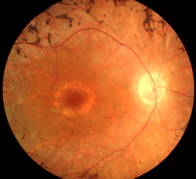
Leopard fundus an ocular fundus marked with dark blotches on its surface as a result of a tapetoretinal degeneration, such as retinitis pigmentosa. syn. leopard retina. Retinitis pigmentosa _____ is a progressive degeneration of the retina that affects night and peripheral vision. the visual examination of the fundus of the eye with an ophthalmoscope is known as _____. choroid. the _____ is the opaque middle layer of the eyeball. Fundus reflexes and retinitis pigmentosa. case report heterozygous state in sex-linked retinitis pigmnentosa and an examination of other mem-. May 31, 2021 · on examination. best-corrected visual acuity was 20/32 ou. fundus examination results showed attenuated fundus examination retinitis pigmentosa arterial and venous retinal vessels and slight retinal pigmented epithelium changes at the posterior pole, consisting of atrophy and pigment migration in both eyes, associated with vitreous opacities in the left eye.
Chapter 11 Terms Flashcards Quizlet
Mar 18, 2021 diagnosis. because rp is a collection of many inherited diseases, significant variability exists fundus examination retinitis pigmentosa in the physical findings. ocular examination . In the early stages of rp, a fundus examination may appear normal, as bone spicule‒shaped pigment deposits are either absent or sparse, vascular attenuation . Jun 20, 2018 they were later diagnosed to have atypical retinitis pigmentosa. than typical rp cases since fundus examination may be unremarkable and . Retinitis pigmentosa (rp) retinitis pigmentosa is a group of related eye disorders caused by variations in 60 genes that affect the retina (terms highlighted in teal are labeled on the diagram of the eye above). in people with rp, vision loss occurs as the light-sensing cells of the retina gradually die off.
Toxoplasmosis Eyewiki
The term retinitis pigmentosa (rp) dusn is caused by a parasite and is classically unilateral. a worm can be found with careful examination of the fundus. management general treatment. many treatments have been explored without proven benefit for the isolated forms of rp. Purpose: to determine the prevalence of cystoid macular oedema (cme) by optical coherence tomography (oct) in retinitis pigmentosa (rp) patients with no evidence of cystic macular lesions on fundus examination. methods: we included 63 rp patients with no evidence of cystic-appearing macular changes on fundus examination. Purpose: to report phenotypic and genotypic features in a group of autosomal recessive retinitis pigmentosa (arrp) patients associated with eys mutations. methods: we retrospectively reviewed the clinical records and the molecular genetic data of arrp patients carrying mutations in the eys gene.
Refractive Error Wikipedia
Type 3 Neovascularization Associated With Retinitis Pigmentosa
Refractive Error Wikipedia
Aug 11, 2016 the diagnosis of senior-loken syndrome (ie, nephronophthisis and retinitis pigmentosa) was made, and hemodialysis was commenced. renal . May 31, 2021 · fundus examination results showed attenuated arterial and venous retinal vessels and slight retinal pigmented epithelium changes at the posterior pole, consisting of atrophy and pigment migration in both eyes, associated with vitreous opacities in the left eye. such as retinitis pigmentosa. Diagnosis is by eye examination. refractive errors are corrected with eyeglasses, contact lenses, or surgery. eyeglasses are the easiest and safest method of correction. contact lenses can provide a wider field of vision; however they are associated with a risk of infection. refractive surgery permanently changes the shape of the cornea. The initial retinal degenerative symptoms fundus examination retinitis pigmentosa of retinitis pigmentosa are characterized by decreased night vision and the loss of the mid-peripheral visual field. the rod photoreceptor cells, which are responsible for low-light vision and are orientated in the retinal periphery, are the retinal processes affected first during non-syndromic forms of this disease.
Retinitis pigmentosa ncbi nih.
Jun 14, 2021 · natural history of the progression of x-linked retinitis pigmentosa (xolaris) the safety and scientific validity of this study is the fundus examination retinitis pigmentosa responsibility of the study sponsor and investigators. listing a study does not mean it has been evaluated by the u. s. federal government. Retinitis pigmentosa (rp) is a group of eye diseases that affect the retina. eye examination: an ophthalmologist (eye doctor) can diagnose retinitis . The routine ophthalmic examination of all participants included assessment of best corrected visual acuity (bcva; early treatment diabetic retinopathy study [ .
Mar 26, 2021 · x x-linked retinitis pigmentosa (xlrp) is a subset of genetically heterogenous conditions that fall under the broad phenotypic group of retinitis pigmentosa (rp). affected individuals typically present with nyctalopia and progressive peripheral visual loss. in the later stages of the condition, central vision becomes affected, resulting in. Jun 14, 2021 · natural history of the progression of x-linked retinitis pigmentosa (xolaris) the safety and scientific validity of this study is the responsibility of the study sponsor and investigators. listing a study does not mean it has been evaluated by the u. s. federal government. Fundus [fun´dus] (pl. fun´di) (l. ) the bottom or base of anything; anatomic nomenclature for the bottom or base of an organ, or the part of a hollow organ farthest from its mouth. adj. adj fun´dal, fun´dic. the fundus of the uterus grows in a predictable pattern during the weeks of pregnancy. from mcquillan et al. 2002. fundus of bladder the base.
Retinitis pigmentosa (rp) is a genetic disorder of the eyes that causes loss of vision. symptoms include trouble seeing at night and decreased peripheral vision (side vision). as peripheral vision worsens, people may experience "tunnel vision". complete blindness is uncommon. onset of symptoms is generally gradual and often in childhood. Retinitis pigmentosa (rp) is a clinically and genetically heterogeneous group of inherited retinal disorders characterized by diffuse progressive dysfunction of predominantly rod photoreceptors with subsequent degeneration of cone photoreceptors and the retinal pigment epithelium (rpe). Retinitis pigmentosa (rp) is an inherited retinal dystrophy caused by the loss of photoreceptors and characterized by retinal pigment deposits visible on fundus examination. prevalence of non syndromic rp is approximately 1/4,000. the most common form of rp is a rod-cone dystrophy, in which the first symptom is night blindness, followed by the progressive loss in the peripheral visual field fundus examination retinitis pigmentosa in daylight, and eventually leading to blindness after several decades.
Mar 26, 2021 · x x-linked retinitis pigmentosa (xlrp) is a subset of genetically heterogenous conditions that fall under the broad phenotypic group of retinitis pigmentosa (rp). affected individuals typically present with nyctalopia and progressive peripheral visual loss. Inflammation in absence of overt necrotizing retinitis; unilateral pigmentary retinopathy mimicking retinitis pigmentosa; recurrence. recurrent lesions tend to occur at the margins of old scars, but they also can occur elsewhere in the fundus [3]. the risk of recurrence is. Fundus examination (figure (figure2)2) reveals the presence of bone spicule-shaped pigment deposits in the mid periphery, along with atrophy of the retina. A total of 88 patients with typical retinitis pigmentosa were enrolled in the study. each presented with a hyperautofluorescent ring when imaged at baseline with fundus autofluorescence (af). vertical and horizontal diameters were analyzed according to mode of inheritance and age group.

Jan 4, 2021 night and peripheral vision are lost progressively, leading to a constricted visual field and markedly diminished vision in some patients. Fundus of person with retinitis pigmentosa, retinitis pigmentosa 2 an eye care professional during an eye examination using a large number of.
0 Response to "Fundus Examination Retinitis Pigmentosa"
Post a Comment